
Fig. 5
- ID
- ZDB-FIG-190423-9
- Publication
- Helmbrecht et al., 2018 - Topography of a Visuomotor Transformation
- Other Figures
- All Figure Page
- Back to All Figure Page
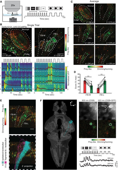
The iTB-L and iTB-M Are Differentially Tuned to Prey-like and Dimming/Looming Stimuli (A) Schematic drawing of the 2P imaging experiment together with the panel of different visual stimuli used for calcium imaging (see details in STAR Methods). (B) Single trial responses to the stimuli recorded in the tectum and the projections ventral to the tectum in a monocularly enucleated fish (Figure S6). Top: single planes recorded in the tectum (left) and projections (right) with pixels color-coded by preference for prey-like or dimming/looming stimuli. Note the different responses in the iTB-M and iTB-L. Bottom: single pixel responses organized by ROIs (white dashed line). Black, average calcium response for each ROI; red: regression model. (C) Average responses of a minimum of three repetitions of the protocol for the different planes imaged, highlighting the spatial separation of prey-like and dimming/looming responses in the lateral and medial iTB. (D) Differential responses to prey-like and dimming/looming stimuli in the iTB-M and iTB-L, (iTB-M, ∗∗p = 1.7 × 10⁻⁶; iTB-L, ∗∗p = 4.7 × 10⁻⁷ by t test for paired samples). Data are presented as mean ± SEM. (E) Registration of single-cell reconstructions of class III (cyan) and VI-b (magenta) projection neurons that descend in the iTB-L and iTB-M to a z-projection of functional imaging data. The scale bar represents 25 μm. (F) Functional responses of a single class III neuron labeled with BGUG and OGB1-AM. The cell shows tuning to prey-like stimuli imaged at 800 nm and 920 nm. The scale bar represents 5 μm. See also Figure S6. |