|
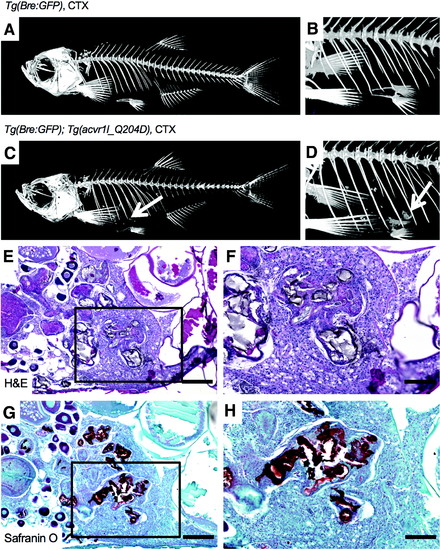
CTX -injected heat-shocked Tg(acvr1l_Q204D) zebrafish exhibited body cavity HO at 8 wpi. Micro-CT imaging of CTX-injected heat-shocked Tg(Bre:GFP); Tg(acvr1l_Q204D-mCherry) zebrafish (C, D) at 8 wpi revealed the presence of HO in the body cavity (arrows in C, D). Control CTX-injected heat-shocked Tg(Bre:GFP) zebrafish did not exhibit detectable HO (A, B). H&E (E, F) and Safranin O (G, H) staining of HO in heat-shocked Tg(Bre:GFP); Tg(acvr1l_Q204D-mCherry) zebrafish revealed heterogeneity of the HO lesion, including numerous proteoglycan-dense regions (G, H, strong red Safranin O stain). (F, H) Enlarged views boxed areas in (E, G), respectively. n = 3 Tg(Bre:GFP); Tg(acvr1l_Q204D-mCherry) zebrafish with CTX treatment. n = 1 Tg(Bre:GFP) zebrafish with CTX treatment. (E, G) 10 × scale bar is 200 μm. (F, H) 20 × scale bar is 100 μm. HO, heterotopic ossification; micro-CT, microcomputed tomography.
|