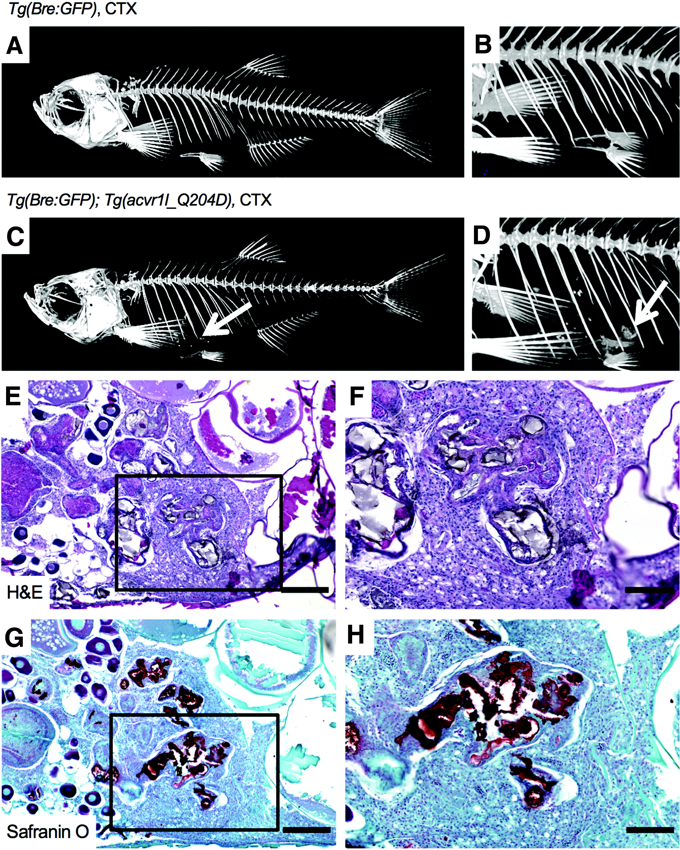
Fig. 4 CTX -injected heat-shocked Tg(acvr1l_Q204D) zebrafish exhibited body cavity HO at 8 wpi. Micro-CT imaging of CTX-injected heat-shocked Tg(Bre:GFP); Tg(acvr1l_Q204D-mCherry) zebrafish (C, D) at 8 wpi revealed the presence of HO in the body cavity (arrows in C, D). Control CTX-injected heat-shocked Tg(Bre:GFP) zebrafish did not exhibit detectable HO (A, B). H&E (E, F) and Safranin O (G, H) staining of HO in heat-shocked Tg(Bre:GFP); Tg(acvr1l_Q204D-mCherry) zebrafish revealed heterogeneity of the HO lesion, including numerous proteoglycan-dense regions (G, H, strong red Safranin O stain). (F, H) Enlarged views boxed areas in (E, G), respectively. n = 3 Tg(Bre:GFP); Tg(acvr1l_Q204D-mCherry) zebrafish with CTX treatment. n = 1 Tg(Bre:GFP) zebrafish with CTX treatment. (E, G) 10 × scale bar is 200 μm. (F, H) 20 × scale bar is 100 μm. HO, heterotopic ossification; micro-CT, microcomputed tomography.
Image
Figure Caption
Figure Data
Acknowledgments
This image is the copyrighted work of the attributed author or publisher, and
ZFIN has permission only to display this image to its users.
Additional permissions should be obtained from the applicable author or publisher of the image.
Full text @ Zebrafish

