
Fig. 1
- ID
- ZDB-FIG-181120-6
- Publication
- Earley et al., 2018 - Critical Role for a Subset of Intestinal Macrophages in Shaping Gut Microbiota in Adult Zebrafish
- Other Figures
- All Figure Page
- Back to All Figure Page
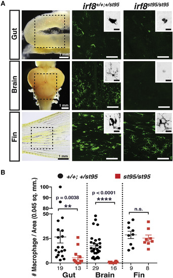
Adult irf8−/− Zebrafish Have a Severe Loss of Tissue-Resident Intestinal and Brain Macrophages but Normal Numbers of Peripheral Macrophages (A) Image analysis was performed on whole-mount gut, brain, and fin dissected from transgenic adult zebrafish expressing the macrophage-specific reporter mpeg1:GFP. Left column shows images of dissected organs and the general region from which several areas were quantified and analyzed (dotted box). Each image represents a quantified 0.045 mm2 field of view. High-magnification insets show an inverted image of a GFP+ macrophage. No clear microglia were detected in irf8 mutant brains. Scale bars in GFP image panels are 50 μm and in insets are 10 μm. (B) Scatterplot shows macrophage number per field of view in each tissue region. Numbers below plot represent the number of areas analyzed from 4 or more animals. Statistical significance was determined by a two-tailed t test. ∗∗p < 0.01, ∗∗∗∗p < 0.0001; n.s., not significant. See also Figure S1. |