
Fig. S5
- ID
- ZDB-FIG-181120-11
- Publication
- Earley et al., 2018 - Critical Role for a Subset of Intestinal Macrophages in Shaping Gut Microbiota in Adult Zebrafish
- Other Figures
- All Figure Page
- Back to All Figure Page
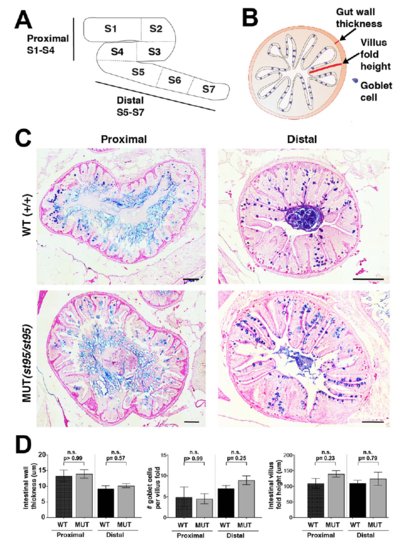
Histological analysis of the intestines does not show structural defects in irf8 mutants, Related to Figure 4. A Schematic of the adult zebrafish intestine and corresponding proximal (S1-S4) and distal (S5-S7) regions. B Schematic of quantified parameters for assessment of intestinal architecture: gut wall thickness, villus fold height, and frequency of goblet cells. C Representative images of proximal (left) and distal (right) intestinal regions stained with combined Alcian Blue and Periodic Acid-Schiff on 5 um transverse sections of irf8 wild type animals (top) and mutant animals (bottom). D Quantification of intestinal wall thickness (left), number of goblet cells per intestinal villus fold (middle) and intestinal villus fold height (right). Data were derived from n= 3 wild type siblings for proximal and distal regions, n=4 mutants for proximal region, and n=5 mutants for distal region. All scale bars represent 100 μm. Statistical significance was determined by the Mann-Whitney test. |