
Fig. S2
- ID
- ZDB-FIG-181030-49
- Publication
- Zimmermann et al., 2018 - Zebrafish Differentially Process Color across Visual Space to Match Natural Scenes
- Other Figures
- All Figure Page
- Back to All Figure Page
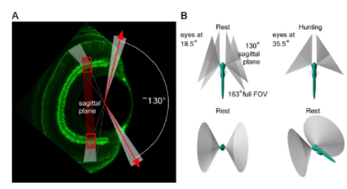
Anisotropic retinal structure. Related to Figure 2. A, Transverse plane (looking down onto the fish from the top) immunolabelled 7 dpf larval zebrafish eye with bipolar cell terminals in green (like Figure 2L) highlights the ~130° visual angle surveyed by the sagittal plane used throughout this work (indicated in red). B, 3D illustrations of the field of view covered by this sagittal plane during “rest” (eyes tilted forward ~18.5°) and during prey-capture (“hunting”, eyes at 35.5°) as indicated. In the top left panel, the ~163° full field of view of the eye is indicated in addition to the 130° field surveyed in this work. |