
Fig. 4
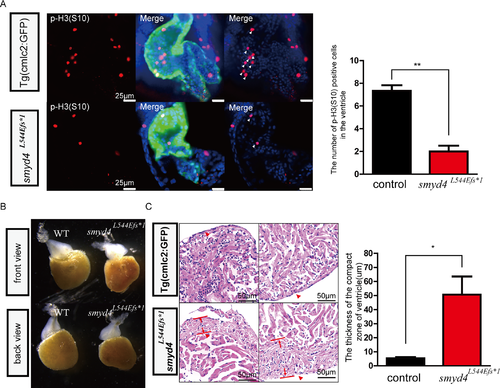
Phenotypical analyses of MZsmyd4L544Efs*1 embryos. (A) The p-H3(S10) immunofluorescence staining showed decreased cell proliferative activity in MZsmyd4L544Efs*1 cardiomyocytes at 48 hpf. The number of p-H3 (S10) positive cells in the MZsmyd4L544Efs*1 ventricles was significantly decreased when compared to the control group (p<0.01, **); (B) The abnormal ventricular morphology of the adult MZsmyd4L544Efs*1 hearts compared to the morphology of control adult hearts; (C) Histological sections and H&E staining images of the adult MZsmyd4L544Efs*1 and control hearts, demonstrating significant thickening of the compact ventricular wall. (The red arrows showed that in the similar compact zone of the ventricle). The quantitative analysis displayed that the thickness of the ventricle compact zone was significantly increased in adult MZsmyd4 L544Efs*1 hearts compared to Tg(cmcl2:GFP) (p<0.05, *). |
| Fish: | |
|---|---|
| Observed In: | |
| Stage: | Long-pec |