
FIGURE
Fig. 6
Fig. 6
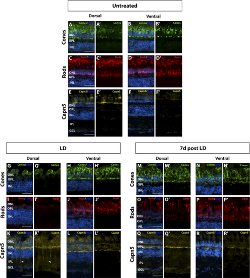
Increase in Capn5 expression correlates with magnitude of retinal damage. (A–B′) Dorsal and ventral expression of cone-specific marker Zpr-1 in undamaged (UT) retina. (C–D′) Dorsal and ventral expression of rod-specific marker 4C12 in the UT retina. (E–F′) IHC for dorsal and ventral Capn5 expression in the UT retina; Capn5 expression is stronger in the dorsal retina (E–E′) compared with the ventral retina (F–F′). (G–H′) Dorsal and ventral cone damage after acute light exposure. There is a significant decrease in the number of cone photoreceptors in the dorsal retina compared with the ventral retina. (I–J′) Dorsal and ventral rod damage after acute light exposure. Rods are almost totally ablated in the dorsal retina, with more moderate damage observed in the ventral retina. (K–L′) IHC for Capn5 expression in the LD retina. Capn5 is strongly upregulated in the INL and surviving cones in the dorsal retina, and more modestly upregulated in the ventral retina. (M–N′) Dorsal and ventral cones have mostly recovered by 7 days post LD. (O–P′) At 7 days post LD, rods are regenerating, with more rods observed in the ventral compared with the dorsal retina. (Q–R′) IHC for Capn5 expression in the 7 days post LD retina. Expression of Capn5 is similar to that of the undamaged retina. Scale bars: 50 μm.
|
Expression Data
| Antibodies: | |
|---|---|
| Fish: | |
| Condition: | |
| Anatomical Terms: | |
| Stage: | Adult |
Expression Detail
Antibody Labeling
Phenotype Data
| Fish: | |
|---|---|
| Condition: | |
| Observed In: | |
| Stage: | Adult |
Phenotype Detail
Acknowledgments
This image is the copyrighted work of the attributed author or publisher, and
ZFIN has permission only to display this image to its users.
Additional permissions should be obtained from the applicable author or publisher of the image.
Full text @ Invest. Ophthalmol. Vis. Sci.