Fig. 3
- ID
- ZDB-FIG-180820-11
- Publication
- Madelaine et al., 2018 - A screen for deeply conserved non-coding GWAS SNPs uncovers a MIR-9-2 functional mutation associated to retinal vasculature defects in human
- Other Figures
- All Figure Page
- Back to All Figure Page
|
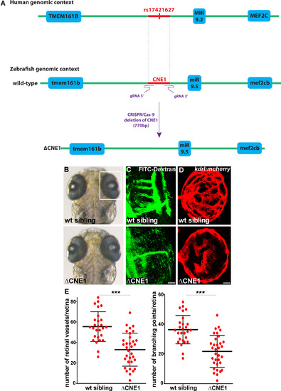
CNE1 regulates retinal vasculature formation in vivo. (A) Schematic of the human and zebrafish genomic DNA containing CNE1, which was deleted using the CRISPR/Cas-9 system with a pair of gRNAs (ΔCNE1 in the zebrafish genome). The deletion is 770 bp long, including the deeply conserved sequence of CNE1 (473 bp; danRer7. chr5:49,928,049–49,928,521) containing the SNP. (B and C) CNE1 homozygous mutants display normal brain and eye morphology (B), but the formation of the hyaloid vasculature in the retina is affected, as revealed by microangiography using FITC-dextran injection (C). The retinal vasculature shown in (C) are from larvae shown in (B). (D) Confocal projections of mCherry immunolabeling in Tg(kdrl:mCherry) retina at 72 hpf showing hyaloid vasculature formation in control and ΔCNE1 mutant larvae. (E) Quantification of the hyaloid vasculature network organization observed in control and homozygous ΔCNE1 mutant larvae at 72 hpf. A minimum of 28 retinas was analyzed for each context. Dorsal view of the brain with anterior up. Lateral view of the retina. Scale bars: 10 μm. Error bars represent s.d. *P < 0.05, **P < 0.001, ***P < 0.0005, determined by t-test, two-tailed. |
| Fish: | |
|---|---|
| Observed In: | |
| Stage: | Protruding-mouth |

