
Fig. 1
- ID
- ZDB-FIG-180817-26
- Publication
- Hofsteen et al., 2018 - ALPK2 Promotes Cardiogenesis in Zebrafish and Human Pluripotent Stem Cells
- Other Figures
- All Figure Page
- Back to All Figure Page
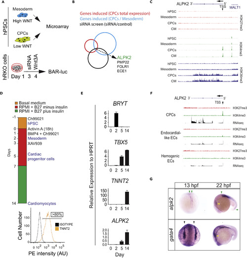
ALPK2 Identification and Expression Analyses (A) Schematic of combinatorial screening of RNA expression from hESC cardiomyocyte differentiation from human embryonic stem cells (hESCs) coupled with an siRNA screen using β-catenin-activated reporter (BAR)-transduced human RKO colon carcinoma cells (hRKO) stimulated with recombinant Wnt3A. (B) Venn diagram identifying Alpha Protein Kinase 2 (ALPK2). (C) Chromatin precipitation followed by deep sequencing (ChIP-Seq) for histone marks H3K4me3, H3K36me3, and H27K4me3 in hESC-derived cultures: hESC (day 0), mesoderm (day 2), cardiac progenitor cells (CPCs, day 5), and cardiomyocytes (day 14) (N = 2). (D) Protocol for high-density monolayer-directed differentiation of hESC-derived cardiomyocytes yielding a high percentage of cardiac troponin T (TNNT2)-positive cells by flow cytometry. (E) Quantitative RT-PCR analysis of markers of mesoderm (Brachyury T, BRYT), cardiac progenitor cells (T-box 5, TBX5), cardiomyocytes (TNNT2), and ALPK2 at days 0, 2, 5, 14 during cardiomyocyte differentiation. (F) RNA sequencing and ChIP-seq for H3K4me3 and H27K4me3 at the ALPK2 locus in CPCs, endocardial-like endothelial cells (EC), and hemogenic ECs (N = 2). (G) In situ hybridization of zebrafish alpk2 and gata4 at 13 hpf and 22 hr post fertilization (hpf, N = 22–36). Green arrowheads denote adaxial cells and primitive somites, black arrowheads mark the bilateral heart fields, and yellow arrowheads denote the primitive heart. Sample size N = 3–5 biological replicates, and data are displayed as mean ± SEM unless otherwise noted. See also Figure S1. |
| Genes: | |
|---|---|
| Fish: | |
| Anatomical Terms: | |
| Stage Range: | 5-9 somites to 26+ somites |