
Fig. 6
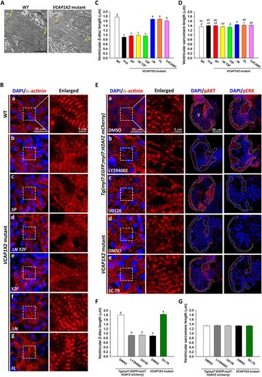
Disorganized sarcomere structure was observed in VCAP1X2 mutant hearts and rescued by VCAP1X2 variant mRNA, hVCAM1 mRNA or a PI3K activator. (A) Transmission electron microscopy shows sparse myofilaments and indistinct Z-discs in ventricular sarcomere of VCAP1X2 mutants at 96 hpf (n = 3 per condition). Arrows indicate Z-discs (Z). Scale bar, 500 nm. (B) Striated Z-discs were detected by α-actinin immunofluorescence in WT heart (a), while a dotted pattern was observed in VCAP1X2 mutants (b) at 96 hpf. ΔN (f) or FL (g), but not SP (c), ΔN Y2F (d) or Y2F (e), restored striated Z-discs (n = 16 per condition, N = 3). Z-disc length and sarcomere length were measured from immunofluorescence images. Z-disc length (C) was reduced in VCAP1X2 mutants and SP, ΔN Y2F or Y2F-injected mutants but not those injected with ΔN, FL, or hVCAM1 mRNA at 96 hpf (n = 160 per condition, N = 3). (D) Slight differences (maximum 8%) were found in sarcomere length, indicating that ventricular sarcomere length was not robustly affected by any treatment at 96 hpf. Data in (C and D) were analyzed by ANOVA with Bonferroni multiple comparisons. Treatments that are not statistically different (α = 0.05) from each other are labeled with the same letter. (E) α-actinin immunofluorescence on sarcomeres of DMSO-treated Tg(myl7:EGFP; myl7:H2AFZ mCherry) embryos (a) or PI3K activator (SC-79)-treated VCAP1X2 mutant heart (e) showed striations, while Tg(myl7:EGFP; myl7:H2AFZ mCherry) embryos treated with inhibitors of PI3K (LY294002) (b) or pMEK (U0126) (c), or DMSO-treated VCAP1X2 mutant hearts (d) showed a dotted pattern. Ventricular myocardium is outlined in right-most panels. Erythrocytes show non-specific staining from secondary antibody. (F) Z-disc length was measured in Tg(myl7:EGFP; myl7:H2AFZ mCherry) embryos and VCAP1X2 mutants treated with PI3K or pMEK inhibitor. Data were analyzed by ANOVA with Bonferroni multiple comparisons (α = 0.05). The difference between groups a and b was significant (p < 0.001). (G) Similar ventricular sarcomere length was detected in all groups (n = 200 per condition, N = 3). ANOVA indicated no significant difference among treatments (p = 0.96). Error bars indicate standard error. |