
Fig. 5
- ID
- ZDB-FIG-180706-9
- Publication
- Siddam et al., 2018 - The RNA-binding protein Celf1 post-transcriptionally regulates p27Kip1 and Dnase2b to control fiber cell nuclear degradation in lens development
- Other Figures
- All Figure Page
- Back to All Figure Page
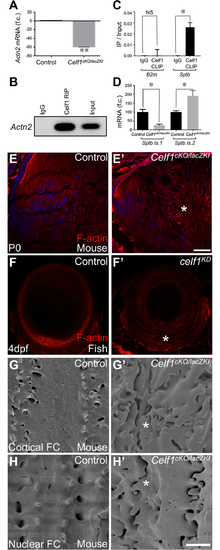
Celf1 deficiency in mouse and fish causes defects in fiber cell morphology. (A) RT-qPCR analysis confirms significant Actn2 down-regulation in Celf1cKO/lacZKI lenses compared to control. (B) RNA immunoprecipitation (RIP) identifies Actn2 as an enriched transcript in Celf1-pulldown in P15 wild-type mouse lens. (C) Cross-linked RNA immunoprecipitation (CLIP) shows Sptb transcripts to be enriched in Celf1-pulldown in wild-type mouse lens. (D) RT-qPCR analysis shows that the high-abundant Sptb isoform (isoform 1 (ENSMUST00000021458)) is reduced, while the low-abundant Sptb isoform (isoform 2 (ENSMUST00000166101)) is abnormally elevated in Celf1cKO/lacZKI lenses. (E, E’) In mouse, phalloidin staining of lens tissue sections (stage P0) shows uniform F-actin deposition along the fiber cell margins in control, while Celf1cKO/lacZKI lenses exhibit abnormal pattern of F-actin (asterisk). (F, F’) In zebrafish, while control lens exhibits normal F-actin deposition, celf1KD lens (stage 4dpf) exhibits abnormal F-actin pattern (asterisk) in fiber cells. (G-H’) In mouse, scanning electron microscopy analysis of cortical and nuclear fiber cells shows disrupted cell organization (asterisk) in Celf1cKO/lacZKI lenses (stage P15). Scale bar in D’ is 75 μm and G’ is 2.5μM. |
| Fish: | |
|---|---|
| Knockdown Reagent: | |
| Observed In: | |
| Stage: | Day 4 |