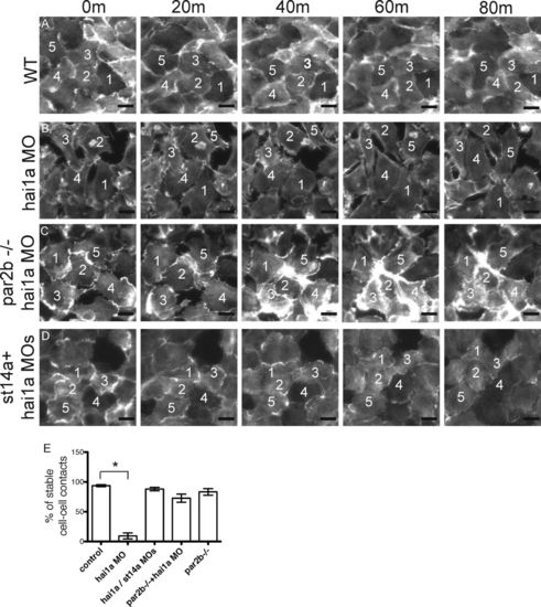
Loss of cell–cell contacts and increased motility of basal layer keratinocytes in hai1a morphants is par2b dependent. (A–D) The yolk sac basal layer of basal-layer-LifeActGFP control or hai1a morphant embryos with or without matriptase or Par2b deficiency was imaged between 24 and 30 hpf. Five panels spanning 80 min (m) are shown. (A) Control embryo. Note tightly apposed basal epithelial cells and stable relative positions of numbered cells. (B) hai1a morphant embryo. Note loss of cell–cell apposition and changing relative cell positions. (C and D) par2b−/− hai1a morphant (C) and st14a/hai1a double morphant (D). Note increased cell apposition and decreased change in position compared with B. Bar, 1 µm. (E) Quantitation of stable cell–cell contacts in control and hai1a morphant basal-layer-LifeActGFP embryos with or without St14a or Par2b deficiency. Time-lapse images of a standard field over the ventral yolk sac were acquired from 24 to 30 hpf for 10 embryos for each condition, and the percentage of individual cells that maintained contact with a neighbor for at least 60 min was determined. Mean ± SEM (n = 10) is shown. Approximately 300 cell–cell contacts were analyzed per condition. The hai1a morphant group was different from controls (*, P < 0.0001), but other groups were not, by one-way ANOVA and Bonferroni posttest.
|
