
Fig. 3
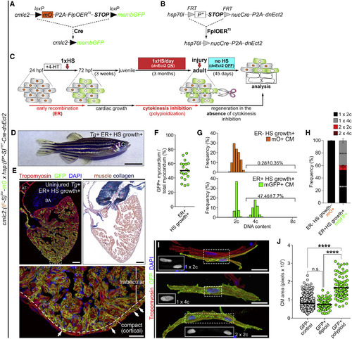
Experimental Strategy to Generate Mosaic Hearts Containing Permanently Labeled Polyploid Cardiomyocytes through Transient Cytokinesis Inhibition (A–C) Transgenes and experimental strategy used to create adult zebrafish with mosaic hearts composed of diploid (GFP−) and polyploid-enriched (GFP+) cardiomyocyte populations. Detailed description of the experimental strategy is provided in Figure S5. (D) External appearance of a double-transgenic adult zebrafish with a mosaic heart composed of GFP− diploid and GFP+ polyploid-enriched cardiomyocytes. (E) Adjacent sections from an adult mosaic heart immunostained for tropomyosin and GFP (left) or stained with acid fuchsin orange G (AFOG) (right). Boxed area shows apex region at higher magnification. White arrows indicate GFP+ cardiomyocytes located in the cortical myocardium. (F) Quantification of the percentage of GFP+ myocardium relative to the total ventricular myocardium from mosaic adult hearts (n = 19). (G) DNA content of GFP− (mO+) and GFP+ cardiomyocytes from mosaic hearts. (H) Quantification of indicated cardiomyocyte populations from (F) (mean ± SD, n = 620 and 520 total cells from 4 to 3 biological replicates per group, respectively; 3 ventricles per replicate; ∗∗∗∗p < 0.0001; ∗∗∗p < 0.001 by one-way ANOVA followed by Tukey's multiple comparisons test). (I) Dissociated cardiomyocytes from a mosaic heart immunostained for tropomyosin and GFP. Boxed regions show DAPI staining. Shown are diploid (top), mononucleated tetraploid (middle), and binucleated tetraploid (bottom) cardiomyocytes. (J) Quantification of cardiomyocyte size from the indicated classes from ventricular dissociations (n = 822, 84, and 97 cardiomyocytes; ∗∗∗∗p < 0.0001; Kruskal-Wallis test followed by Dunn's multiple comparisons test). CM, cardiomyocyte; ER, early recombination; HS, heat-shocked. Scale bars, 5 mm (D), 100 μm (E), and 25 μm (I). |
| Gene: | |
|---|---|
| Fish: | |
| Condition: | |
| Anatomical Term: | |
| Stage: | Adult |
| Fish: | |
|---|---|
| Condition: | |
| Observed In: | |
| Stage: | Adult |
Reprinted from Developmental Cell, 44, González-Rosa, J.M., Sharpe, M., Field, D., Soonpaa, M.H., Field, L.J., Burns, C.E., Burns, C.G., Myocardial Polyploidization Creates a Barrier to Heart Regeneration in Zebrafish, 433-446.e7, Copyright (2018) with permission from Elsevier. Full text @ Dev. Cell