
Fig. 10
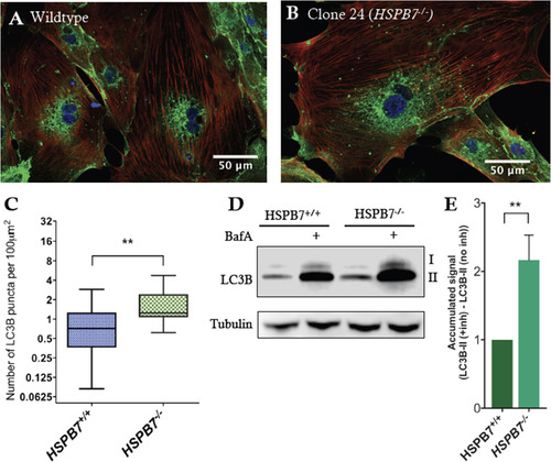
Upregulation of autophagic processes in HSPB7 null cardiomyocytes. Shown are representative images of immunofluorescence for LC3B (green) in (A) wildtype or (B) HSPB7 mutant hESC-derived cardiomyocytes. Samples are co-stained with anti-cardiac troponin T (red) and counterstained with DAPI (blue). (C) Quantification of LC3B puncta in hESC-CMs showing an increase in average number of puncta in HSPB7 mutant cardiomyocytes. (D) Western blot for LC3B in the presence or absence of autophagy inhibitor bafilomycin A (BafA). (E) Quantification of LC3BII western blot by densitometry reveals an increase in the accumulation of LC3BII in HSPB7-/- hESC-CMs during bafilomycin A treatment when compared with HSPB7 wildtype controls. For C and E, changes are significant according to an unpaired t-test, corrected for multiple comparisons with the Holm-Sidak method. |
Reprinted from Developmental Biology, 435(1), Mercer, E.J., Lin, Y.F., Cohen-Gould, L., Evans, T., Hspb7 is a Cardioprotective Chaperone Facilitating Sarcomeric Proteostasis, 41-55, Copyright (2018) with permission from Elsevier. Full text @ Dev. Biol.