
Fig. 1
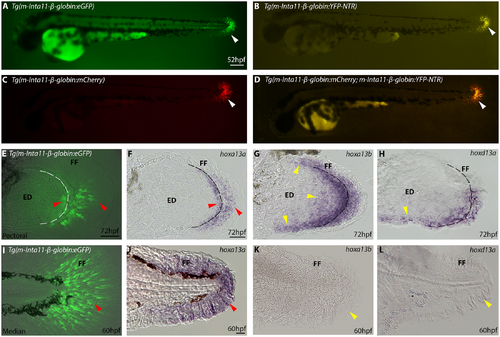
Nitroreductase (NTR) in Tg(Inta11:NTR) is expressed in subset of hoxa13a/hoxd13a-expressing mesenchyme of pectoral fin fold and hoxa13a-only expressing mesenchyme of the median fin fold. (A-D) Whole mount view of transgenic lines using the “m-Inta11-β-globin” regulatory elements at 52hpf. (E-H) Pectoral fin dissections showing reporter, hoxa13a, hoxa13b, and hoxd13a expression in the fin fold mesenchyme at 72hpf. (I-L) Median fin dissections showing reporter, hoxa13a, hoxa13b, and hoxd13a expression in the fin fold mesenchyme at 60hpf. At 52hpf, transgene (eGFP, mCherry, YFP-NTR) expression is visible in the migrating mesenchyme of the median fin fold using the “m-Inta11-β-globin” regulatory elements (white arrow) (A-D). Double transgenic fish Tg(m-Inta11-β-globin:mCherry; m-Inta11-β-globin:YFP-NTR) show colocalization of mCherry and YFP expressing cells in the median fin fold (D). Reporter expression is present in the migrating mesenchyme within the pectoral fin fold, as well as cells located at the distal edge of the endoskeletal disc (red arrow) (E), recapitulating a subset of hoxa13a/hoxd13a-expressing cells (F). hoxa13b, and hoxd13a expression extends proximally within the endoskeletal disk and this region does not correlate with reporter expression (yellow arrows) (G, H). Dotted line represents limit between fin fold and endoskeletal disc (F-H). Reporter expression is present in the migrating mesenchyme within the median fin fold (red arrow) (I), recapitulating endogenous hoxa13a expression (J). No hoxa13b or hoxd13a expression is visible in the median fin at 60hpf (yellow arrows) (K, L). Brightfield (F-H, J-L), fluorescence (A-D), and brightfield/fluorescence merged images (E, I K). ED, Endoskeletal disc; FF, Fin fold. Scale bars: 200μm in A-D; 50μm in E-G, I-L; 30μm in H. |