
Fig. S1
- ID
- ZDB-FIG-180529-44
- Publication
- Bertuzzi et al., 2018 - Spinal cholinergic interneurons differentially control motoneuron excitability and alter the locomotor network operational range
- Other Figures
- All Figure Page
- Back to All Figure Page
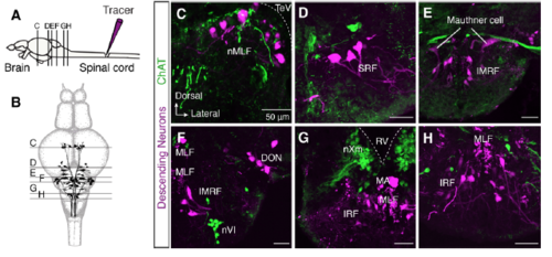
Brain neurons descending to spinal cord are not cholinergic. (A-B) Injection of methylrhodamine dextran in the spinal cord retrogradely labels all the descending supra-spinal neurons (N = 8 zebrafish brains). Schematic representation of the distribution of adult zebrafish brain descending neurons with the level of sections that correspond to the following images in C-H. (C-H) Confocal images show that none of the descending labeled neuron is ChAT+ in all studied brain areas. DON, descending octaval nucleus; IMRF, intermediate reticular formation; IRF, inferior reticular formation; MA, Mauthner axon; MLF, medial longitudinal fascicle; nMLF, nucleus of the medial longitudinal fascicle; nVI, abducens nucleus; nXm, vagal motor nucleus; RV, rhombencephalic ventricle; SRF, superior reticular formation; TeV, tectal ventricle. |