
Fig. S3
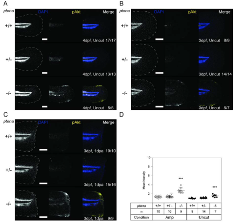
Elevated p-AKT in the caudal fin-folds of Pten deficient embryos. (A) Uncut embryos from a ptena+/-ptenb-/- in-cross were fixed at 4dpf (4dpf, uncut) in parallel to the embryos depicted in Fig. 2A. (B, C) Embryos from a ptena+/-ptenb-/- in-cross were amputated and fixed at 1dpa (i.e. 3dpf, 1dpa), equivalent uncut controls (3dpf, uncut) were included. Embryos were subjected to whole-mount immunohistochemistry using a p-AKT-specific antibody (p-S473) (yellow). The embryos were counterstained with DAPI (blue). Representative images of embryo caudal fin-folds are shown, and in the left panels the edge of the fin-fold is indicated with a dashed line. Number of embryos showing similar patterns/ total number of embryos analysed is indicated in the bottom right corner. The scale bar represents 100μm. (D) p-AKT immunofluorescence was quantified by mean intensity of the region between the notochord and the edge of the caudal fin-fold. Means within amputated or uncut groups are compared to ptena+/+ptenb-/- embryos. The number of embryos analysed is indicated (n). Significance: *** p<0.001; error bars represent standard deviation. Quantification of p-AKT immunofluorescence at 4dpf is depicted in Fig. 2B. |