
FIGURE
Fig. 6
Fig. 6
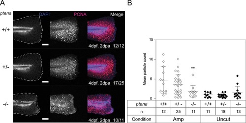
Arrested proliferation at the edge of the amputated caudal fin‐fold of Pten deficient embryos by 2 dpa. (A) Embryos from a ptena+/−ptenb−/− in‐cross were fixed at 2 dpa (i.e., 4 dpf, 2 dpa) and subjected to whole‐mount immunohistochemistry using an antibody specific for PCNA (red). The embryos were counterstained with DAPI (blue). Maximum intensity projection images were taken of the caudal fin‐folds and all embryos were genotyped. Representative images of amputated embryo caudal fin‐folds are shown and the number of embryos showing similar patterns/total number of embryos analyzed is indicated in the bottom right corner of the right panels. The scale bar represents 100 μm. (B) PCNA immunofluorescence between the tip of the notochord and edge of the caudal fin‐fold was quantified by mean particle count. Means within amputated or uncut groups were compared to ptena+/+ptenb−/− embryos. Significance: **p < 0.01; error bars represent standard deviation
|
Expression Data
Expression Detail
Antibody Labeling
Phenotype Data
Phenotype Detail
Acknowledgments
This image is the copyrighted work of the attributed author or publisher, and
ZFIN has permission only to display this image to its users.
Additional permissions should be obtained from the applicable author or publisher of the image.
Full text @ Regeneration (Oxf)