
FIGURE
Fig. 6
- ID
- ZDB-FIG-180510-30
- Publication
- Wiggenhauser et al., 2017 - Studying Diabetes Through the Eyes of a Fish: Microdissection, Visualization, and Analysis of the Adult tg(fli:EGFP) Zebrafish Retinal Vasculature
- Other Figures
- All Figure Page
- Back to All Figure Page
Fig. 6
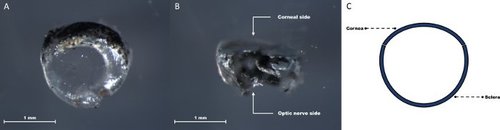
Corneosclera consisting of sclera and cornea, which is disconnected from the remaining intraocular tissue. Corneal side view showing the continuation of the translucent cornea into the pigmented sclera (A). Lateral view focused on the scleral part of the corneosclera (B). Schematic depiction of the removed corneosclera (C). |
Expression Data
Expression Detail
Antibody Labeling
Phenotype Data
Phenotype Detail
Acknowledgments
This image is the copyrighted work of the attributed author or publisher, and
ZFIN has permission only to display this image to its users.
Additional permissions should be obtained from the applicable author or publisher of the image.
Full text @ J. Vis. Exp.