
FIGURE
Fig. 4
- ID
- ZDB-FIG-180510-28
- Publication
- Wiggenhauser et al., 2017 - Studying Diabetes Through the Eyes of a Fish: Microdissection, Visualization, and Analysis of the Adult tg(fli:EGFP) Zebrafish Retinal Vasculature
- Other Figures
- All Figure Page
- Back to All Figure Page
Fig. 4
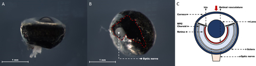
Adult zebrafish eye after removal of all extraocular muscles. Lateral view with the cleared outer rim of the ocular globe visible (A). Optic nerve side view with optic nerve in the middle. The light-reflecting sclera is not covering the whole area around the nerve (red dashed line) (B). Schematic depiction of the zebrafish eye at this step (C). |
Expression Data
Expression Detail
Antibody Labeling
Phenotype Data
Phenotype Detail
Acknowledgments
This image is the copyrighted work of the attributed author or publisher, and
ZFIN has permission only to display this image to its users.
Additional permissions should be obtained from the applicable author or publisher of the image.
Full text @ J. Vis. Exp.