
Fig. 1-S8
- ID
- ZDB-FIG-180504-16
- Publication
- Weber et al., 2017 - Cell-accurate optical mapping across the entire developing heart
- Other Figures
- All Figure Page
- Back to All Figure Page
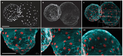
Myocardial morphology is heterogeneous across the heart at 52 hpf. Maximum intensity projections of point-scanning confocal and two-photon microscopy z-stacks recorded from a transgenic zebrafish embryo expressing myl7:H2A-mCherry and myl7:lck-EGFP in the myocardium. Scale bars: 50 µm. (a) Positions of myocardial nuclei (myl7:H2A-mCherry) across the heart. (b) Myocardial cell membranes (myl7:lck-EGFP). (c) Color overlay of cell membranes (cyan) and nuclei (red). (c1-3) Detailed view (rotated by −15 degrees about x–axis) of the cell morphologies in inflow, inner curvature and outer curvature regions of the atrium, as indicated in panel (c). |