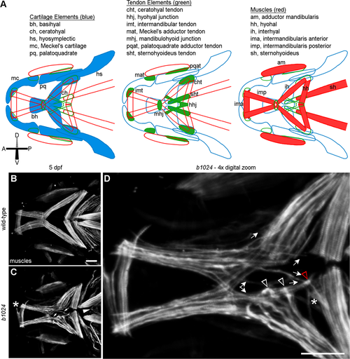
Homozygous carriers of b1024 display second pharyngeal arch lower jaw muscle defects. (A) Schematic of connective tissues in the larval zebrafish jaw, modified from Chen and Galloway, 2014. (B, C) Ventral craniofacial muscles in wild-type and b1024 embryos. (C) Muscle fibers from an intermandibularis posterior muscle extend past the mandibulohyoid junction and insert alongside the adjacent interhyal muscle or at the hyohyal junction (arrows). Muscle fibers apparently from the sternohyoideus are present in the ventral midline of mutants (arrowhead). Ectopic muscle fibers split off from the intermandibularis anterior muscle in a small percentage of mutants (asterisk). (D) 4x digital zoom of a single Z slice from confocal image in C. Arrows show ectopic paths of intermandibularis posterior muscle fibers. Arrowheads show ectopic midline attachments of interhyal muscle fibers. A three-way junction (red arrowhead in D) forms between ectopic intermandibularis posterior, interhyal, and hyohyal (asterisk in D) muscle fibers. All images ventral view, anterior to the left. Scale bars = 50 μm
|
