
Fig. S1
- ID
- ZDB-FIG-180420-15
- Publication
- Kawasaki et al., 2017 - Development and growth of organs in living whole embryo and larval grafts in zebrafish
- Other Figures
- All Figure Page
- Back to All Figure Page
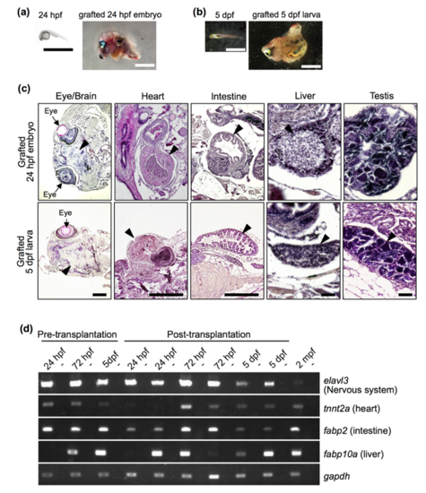
Development of grafted 24 hpf embryos and 5 dpf larvae at 2 months post-transplantation. (A, B) Morphology of 24 hpf embryos (A) and 5 dpf larvae (B) before and after transplantation. Scale bar: 2 mm. (C) Histology of grafted 24 hpf embryos and 5 dpf larvae. Sections were processed with haematoxylin and eosin staining, and representative major organs are shown. Note that morphologically distinct organs developed from 24 hpf embryos and 5 dpf larvae. Arrowheads indicate each organ, as labelled above the panels. Photos of the heart, liver and testis were from grafts in which those organs developed. Scale bars: 500 µm (brain, heart and intestine) and 50 µm (liver and testis). (D) RT-PCR analysis of organ-specific genes in grafted 24 hpf and 72 hpf embryos and 5 dpf larvae at 2 months post-transplantation. Duplicate experiments were performed with independent samples. Wild-type zebrafish at 2 mpf was used as a positive control. Negative controls lacking reverse transcriptase are shown in alternating lanes (-). gapdh was used as a positive control. |