
Fig. 5
- ID
- ZDB-FIG-180420-14
- Publication
- Kawasaki et al., 2017 - Development and growth of organs in living whole embryo and larval grafts in zebrafish
- Other Figures
- All Figure Page
- Back to All Figure Page
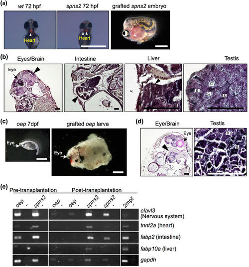
Development of lethal mutant embryos and larvae grafted into rag1 mutants. (a,c) Morphology of 72 hpf spns2 mutant embryos (a) and 7 dpf oep mutant larvae (c) before and after transplantation into germ cell-depleted rag1 mutants. The spns2 mutant has cardia bifida (two hearts) as determined by cmlc2-driven mRFP expression (arrowheads), and the oep mutant exhibits a single eye (arrows). Scale bar: 1 mm. (b,d) Histology of organs found in grafted spns2 mutant embryos (b) and oep mutant larvae (d) at 2 months after transplantation. Arrowheads indicate each organ as labelled above the panels. Note that testes of the grafted spns2 and oep mutants contained all stages of spermatogenic cells: spermatogonia (g), spermatocytes (c), and sperm (s). The heart was not observed in grafted spns2 mutant embryos (b). Scale bar: 200 µm. (e) RT-PCR analysis of grafted spns2 and oep mutants at 2 months post-transplantation. Wild-type zebrafish at 2 mpf were used as a positive control. Negative controls lacking reverse transcriptase are in alternating lanes (−). Each experiment was conducted using two independent samples. |