
FIGURE
Fig. 1
- ID
- ZDB-FIG-180411-5
- Publication
- Veil et al., 2017 - Maternal Nanog is critical for the zebrafish embryo architecture and for cell viability during gastrulation
- Other Figures
- All Figure Page
- Back to All Figure Page
Fig. 1
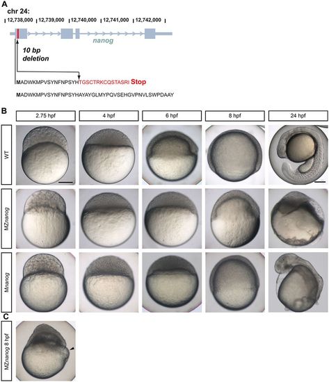
Live phenotypes of Nanog mutants. (A) Scheme of the introduced mutation in the zebrafish nanog gene by TALENs. In the first exon 10 bp were deleted (arrow). Amino acids changed in the nanog mutant are shown in red. The wild-type Nanog amino acid sequence is shown beneath. (B) Lateral view of wild-type (WT), MZnanog and Mnanog embryos from 2.75 to 24 hpf. (C) Blastoderm detachment and start of yolk lysis (arrowhead) in an MZnanog embryo. Scale bar: 200 µm. |
Expression Data
Expression Detail
Antibody Labeling
Phenotype Data
| Fish: | |
|---|---|
| Observed In: | |
| Stage Range: | Sphere to Prim-5 |
Phenotype Detail
Acknowledgments
This image is the copyrighted work of the attributed author or publisher, and
ZFIN has permission only to display this image to its users.
Additional permissions should be obtained from the applicable author or publisher of the image.
Full text @ Development