
FIGURE
Fig. S1
- ID
- ZDB-FIG-180411-4
- Publication
- Mateos et al., 2017 - Correlative Super-resolution and Electron Microscopy to Resolve Protein Localization in Zebrafish Retina
- Other Figures
- All Figure Page
- Back to All Figure Page
Fig. S1
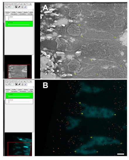
Alignment of light and electron microscopy images. A. Screenshot from TrackEM2 interface with SEM image and numbered landmarks (yellow) along different nuclei. B. Screenshot from TrackEM2 interface with fluorescence image and numbered landmarks (yellow) along different DAPI stained nuclei. To change layer transparency the sliders on the left upper part of the menu can be used. Scale bar: 1 µm. |
Expression Data
Expression Detail
Antibody Labeling
Phenotype Data
Phenotype Detail
Acknowledgments
This image is the copyrighted work of the attributed author or publisher, and
ZFIN has permission only to display this image to its users.
Additional permissions should be obtained from the applicable author or publisher of the image.
Full text @ J. Vis. Exp.