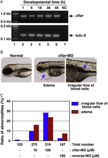
Developmental anomalies in zebrafish embryos with knockdown of cflar transcripts. (A) The expression profile of zebrafish cflar transcripts during embryogenesis. Total RNAs isolated from embryos, which were collected at 1, 6, 18, 36, and 48 h after fertilization (lanes 2–6), was analyzed by RT-PCR. PCR products amplified with primers specific for cflar (upper panel) and tubulin α6 (tubα6) (lower panel) were resolved by agarose-gel electrophoresis. Molecular weight markers were run in lane 1, and a negative control (NC) with no template DNA was run in lane 7. Arrows indicate the expected positions of the cflar and tubα6 PCR products, respectively. (B) Morphological analysis of developing embryos subjected to morpholino oligonucleotide (MO) injection. Fertilized eggs were injected with an antisense MO for cflar, cflar-MO and their development was monitored under the microscope. Images of the developing embryos were acquired at three days later. The resulting abnormal phenotypes consisted of edema (middle panel) and a cluster of blood cells in the vessel (right panel), indicated by an arrow. (C) A summary of the phenotypic data presented in (B). Embryos displaying the edema (red) or irregular flow of red blood cells (blue) were counted under the microscope. Data represent the percentages calculated from four independent experiments.
|
