
Fig. 4
- ID
- ZDB-FIG-180403-13
- Publication
- Posner et al., 2017 - The zebrafish as a model system for analyzing mammalian and native α-crystallin promoter function.
- Other Figures
- All Figure Page
- Back to All Figure Page
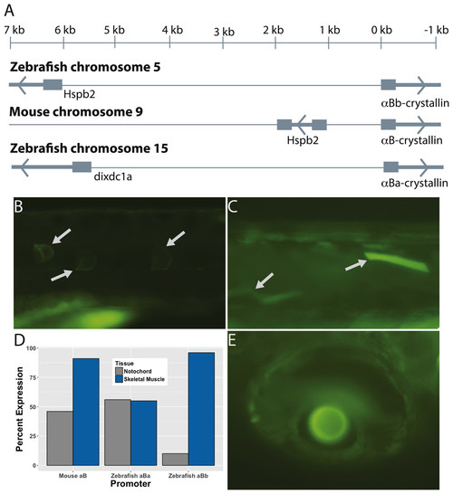
The paralogous zebrafish αBa- and αBb-crystallin promoters produced similar, but distinct, GFP expression profiles. Zebrafish αBb-crystallin has the same syntenic relationship with Hspb2 as mouse αB-crystallin, although the intergenic region between the two genes is much larger in the zebrafish (A). The zebrafish αBa-crystallin paralog has moved to a separate chromosome. Both zebrafish paralogs produced GFP expression most often in notochord (B) and skeletal muscle (C). The αBa paralog drove expression in these tissues equally while αBb was more active in skeletal muscle (D). Expression in lens (E) and extralenticular regions of the eye was more rare. Images shown are representative with the details of GFP expression not differing noticeably between paralogs or the promoter length used. |