
Fig. 3
- ID
- ZDB-FIG-180321-14
- Publication
- Campbell et al., 2017 - Phosphodiesterase Inhibitors Sildenafil and Vardenafil Reduce Zebrafish Rod Photoreceptor Outer Segment Shedding
- Other Figures
- All Figure Page
- Back to All Figure Page
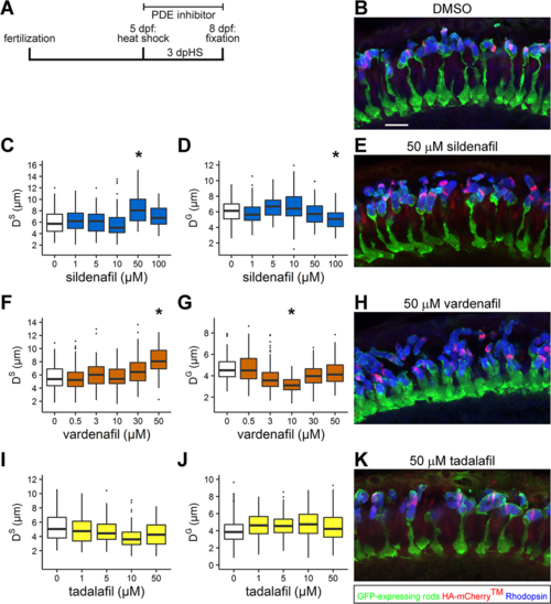
Sildenafil and vardenafil reduce larval rod outer segment shedding and vardenafil changes rod photoreceptor morphology. (A) Schematic timeline for examining rod outer segment renewal in 8 dpf, 3 days post heat shock (dpHS) Tg(Xla.rho:EGFP); Tg(hsp70:HA-mCherryTM); albb4/b4 fish after bathing in phosphodiesterase inhibitor. (B, E, H, K) Representative images of photoreceptor layers from 8 dpf, 3 dpHS larvae treated with (B) 0.05% DMSO, (E) 50 μM sildenafil, (H) 50 μM vardenafil, and (K) 50 μM tadalafil. Retinal sections were immunolabeled with anti-GFP (green), anti-HA (red), and anti-Rhodopsin (blue) antibodies. Images are projections of a subset of z-sections totaling 5.97 μm. Scale bar: 10 μm. Quantification of (C, F, I) shedding distance (DS) and (D, G, J) growth distance (DG). Lower and upper hinges of box correspond to first and third quartiles; middle corresponds to median; whiskers extend 1.5 * interquartile range above and below the hinges; dots represent outliers. Graphs represent (C, D) sildenafil n = 5 fish/treatment ([C] ≥63 and [D] ≥93 outer segments); (F, G) vardenafil n = 4 fish/treatment ([F]) ≥91 and [G] ≥122 outer segments); and (I, J) tadalafil n = 4 fish/treatment except for the 10 μM tadalafil sample with n = 3 fish ([I] ≥100 and [J] ≥190 outer segments). *95% CI of difference compared to control does not span zero. |