
Fig. 7
- ID
- ZDB-FIG-180223-6
- Publication
- Meng et al., 2017 - AIBP Limits Angiogenesis Through γ-Secretase-Mediated Upregulation of Notch Signaling
- Other Figures
- All Figure Page
- Back to All Figure Page
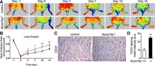
Laser Doppler Blood Perfusion (LDBP) imaging of control and AIBP (apolipoprotein A-I [apoA-I]–binding protein) knockout (KO) mice. A, Mice were subjected to hindlimb ischemia, and representative Laser Doppler images were shown at the indicated time points. Arrows point to the ischemic limbs. B, Quantitative assessment of limb perfusion. The results from AIBP KO mice are shown in red. C, Immunohistochemistry (IHC) analysis of CD31 (cluster of differentiation 31 protein) staining in the ischemic tissue from control or AIBP KO mice. D, Quantification of CD31 staining in C. Scale bar: 50 μm. Mean±SE; n=8 in each group, *P<0.05; **P<0.01. The statistical analysis was done using 1-way analysis of variance (ANOVA). |