
FIGURE
Fig. 3
- ID
- ZDB-FIG-180130-24
- Publication
- Morris et al., 2017 - Transmission Electron Microscopy of Zebrafish Spinal Motor Nerve Roots
- Other Figures
- All Figure Page
- Back to All Figure Page
Fig. 3
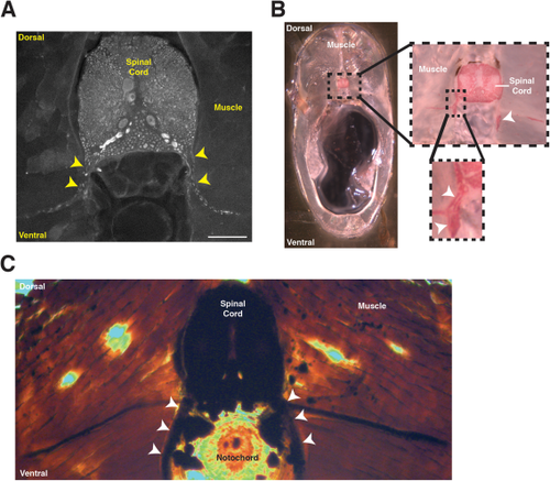
Locating spinal motor nerves in adult zebrafish transverse sections. A: Immunohistochemistry on floating sections using an antibody to tubulin. Yellow arrowheads identify the tubulin+ peripheral motor nerves. Scale bar = 100 µm. B: Black Gold II myelin stain to label the myelin sheath. The dotted box denotes the area enlarged in the right and bottom right images. White arrowheads identify the myelin sheath associated with peripheral motor axons. C: Osmium labeling identifies the myelin sheath associated with peripheral motor axons (white arrowheads). |
Expression Data
Expression Detail
Antibody Labeling
Phenotype Data
Phenotype Detail
Acknowledgments
This image is the copyrighted work of the attributed author or publisher, and
ZFIN has permission only to display this image to its users.
Additional permissions should be obtained from the applicable author or publisher of the image.
Full text @ Dev. Dyn.