
Fig. 6
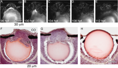
Formation of the second lens cell mass in occ. 2-photon live-embryo images of an occ eye were compared with 10-μm frozen sections stained with H&E of occ and wt eyes. The cornea is at the top of each image and the retina is at the bottom. The lens epithelial cells were observed in the developing lens in occ eyes from 4 to 6 dpf (A–E). In the occ eye, the lens epithelial cells were multilayered and formed a cell mass secondary to the original lens (F,G). In the wt eye, the lens epithelial cells formed a single layer inside of the lens capsule (H). CO, cornea; LE, lens epithelial cells. Scale bar = 30 μm for A–E, and = 20 μm for F–H. |
| Fish: | |
|---|---|
| Observed In: | |
| Stage: | Day 4 |