
FIGURE
Fig. 4
Fig. 4
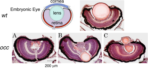
Although two lens masses formed in the occ eye, the retina appeared normal. The layered structure of the retina in the occ eye was similar in size to that of the wt. In the wt eye, the lens was large and spherically symmetric. In the occ eye, the primary lens remained intact and was displaced posteriorly, pushing into the developing retina (10-μm frozen sections were stained with H&E at 5 dpf. Scale bar = 200 μm.). The drawing of the embryonic eye is from Soules and Link, 2005 |
Expression Data
Expression Detail
Antibody Labeling
Phenotype Data
| Fish: | |
|---|---|
| Observed In: | |
| Stage: | Day 5 |
Phenotype Detail
Acknowledgments
This image is the copyrighted work of the attributed author or publisher, and
ZFIN has permission only to display this image to its users.
Additional permissions should be obtained from the applicable author or publisher of the image.
Full text @ Dev. Dyn.