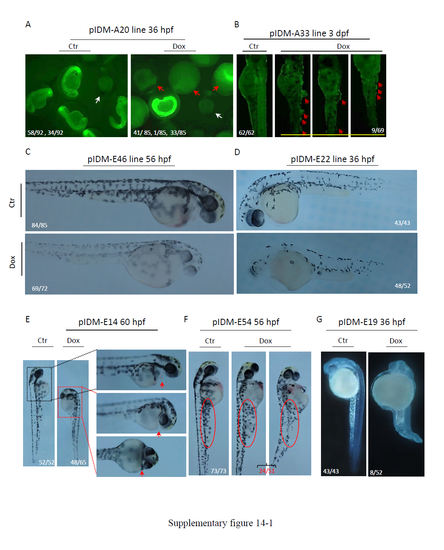
Fig. S14
- ID
- ZDB-FIG-180126-53
- Publication
- Ma et al., 2017 - A novel inducible mutagenesis screen enables to isolate and clone both embryonic and adult zebrafish mutants
- Other Figures
- All Figure Page
- Back to All Figure Page
|
Morphology of Dox treated pIDM mutants with multiple insertions. (A) Line pIDM-A20. Early embryonic lethality for Dox treated pIDM-A20 embryos at 36 hpf. Red arrow, dead embryos; white arrow, WT embryos. In untreated pIDM-A20 embryos, 58 embryos were EGFP positive and 34 embryos were EGFP negative. None of these embryos had died. In Dox treated pIDM-A20 embryos (total 85), 41 embryos were dead at 36 hpf, while among 34 living embryos, only one embryo (with clearly retarded development) was EGFP positive with 33 embryos EGFP negative. (B) Line pIDM-A33. Epidermal blisters in Dox treated pIDM-A33 embryos at 3 dpf. Red arrow, lesions. (C) Line pIDM-E46. Less pigmentation in Dox treated pIDM-E46 embryos at 56 hpf. (D) Line pIDM-E22. Less pigmentation in Dox treated pIDM-E22 embryos at 36 hpf. (E) Line pIDM-E14. Short stature and no pericardium in Dox treated pIDM-E14 embryos at 60 hpf. Red arrow, pericardium. (F) Line pIDM-E54. Shorter and thicker yolk extension in Dox treated pIDM-E54 embryos at 56 hpf. Red ellipse, yolk extension. (G) Line pIDM-E19. Small head, curved body and unabsorbed yolk in Dox treated pIDM-E19 embryos at 36 hpf. (H) Line pIDM-A199. Curved body with severe cell death in Dox treated pIDM-A199 embryos at 36 hpf. (I) Line pIDM-E256. Arrested development in Dox treated pIDM-E256 embryos at 30 hpf. (J) Line pIDM-A96. Arrested development in Dox treated pIDM-A96 embryos at 30 hpf. (K) Line pIDM-A28. Short stature in Dox treated pIDM-A28 embryos at 60 hpf. (L) Line pIDM-E1. Pericardial edema and deformed head in Dox treated pIDM-E1 embryos at 36 hpf. Red arrow, pericardial edema; (*), deformed head. Number in B-L, type of represented embryos/total embryos in F2 population. Details of insertion positions, orientation of pIDM and expression changes of tagged genes in each line are provided in Table 1. |


