
Fig. 2
- ID
- ZDB-FIG-180109-19
- Publication
- Niklaus et al., 2017 - Shaping of Signal Transmission at the Photoreceptor Synapse by EAAT2 Glutamate Transporters
- Other Figures
- All Figure Page
- Back to All Figure Page
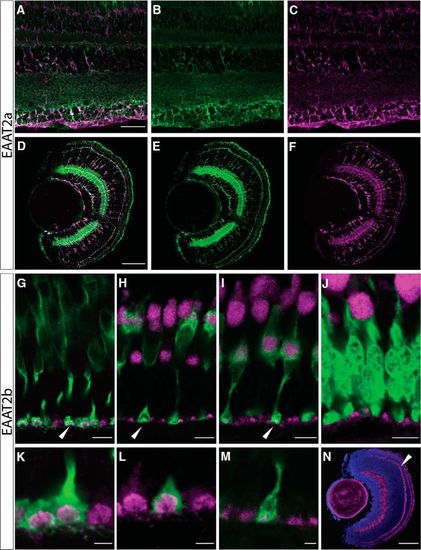
Protein expression of EAAT2 paralogs. A–F, Double immunostaining of EAAT2a (green) and glutamine synthetase (magenta) in adult (A) and larval (5 dpf; D) retinal sections confirm expression of EAAT2a in Müller glia cells. Separated channels are shown in B (adult), E (5 dpf; EAAT2a, green channel only), C (adult), and F (5 dpf; glutamine synthetase, magenta channel only). Scale bar in A is 30 µm; also applies to B and C. Scale bar in D is 50 µm; also applies to E and F. G–N, EAAT2b protein is expressed in a dotted manner in the outer plexiform layer (OPL) in all cone pedicles, but it is not expressed in rods. EAAT2b antibody staining (magenta) on adult retinal sections stained with Zpr-1 (red-green double cones, G) and on retinal sections of zebrafish expressing GFP in blue cones (H), UV cones (I), and rods (J) confirms that EAAT2b is cone specific and is spared from rod spherules. K–M show zoom-ins of the cone pedicles expressing EAAT2b (magenta) in red-green double cones (K), blue cones (L), and UV cones (M). N shows larval (5 dpf) expression of EAAT2b in magenta together with a nuclear counterstain (DAPI, blue). Scale bars in G–J are 7 µm. Scale bars in K–M are 2 µm. Scale bar in N is 30 µm. |
| Genes: | |
|---|---|
| Antibodies: | |
| Fish: | |
| Anatomical Terms: | |
| Stage Range: | Day 5 to Adult |