
Fig. 4
- ID
- ZDB-FIG-180108-13
- Publication
- Bremer et al., 2017 - A small molecule screen identifies in vivo modulators of peripheral nerve regeneration in zebrafish
- Other Figures
- All Figure Page
- Back to All Figure Page
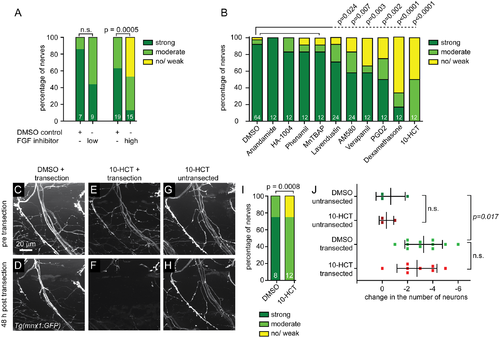
FGF inhibitor and six additional compounds that impair nerve regeneration after fin removal also impair regeneration after laser nerve transection. (A) Ventral nerves of Tg(mnx1:GFP) transgenic larvae were laser transected and treated with DMSO for control, or treated with a low (17μM) or high (34μM) dose of the FGF inhibitor SU5402. Regeneration was scored 48 hours later and the graphical representation of the extent of nerve regeneration (no/ weak, moderate, strong regeneration) is shown, demonstrating that the FGF inhibitor SU5402 significantly and dose-dependently impairs nerve regeneration. (B) Quantification of dorsal nerve regeneration in Tg(isl1:GFP) transgenic larvae measuring the extent of dorsal nerve regeneration (no/ weak, moderate, strong regeneration) for controls (0.5% DMSO) and all tested compounds. Post nerve transection exposure to 100μM anandamide (cannabinoid agonist), 25μg/ml HA-1004 (PKC inhibitor), 10μg/ml phenamil (TRPP3 channel blocker), and 25μg/ml MnTBAP (SOD mimetic) did not significantly impair nerve regeneration. However, 25μg/ml lavendustin (EGFR inhibitor), 4μM AM-580 (RA receptor agonist), 10μg/ml verapamil (calcium channel blocker), 5μM PGD2, 25μg/ml dexamethasone (corticosteroid), and 250μM 10-HCT significantly impaired nerve regeneration. (C-I) Ventral nerves of Tg(mnx1:GFP) transgenic larvae treated with DMSO for control (C, D), or with 125μM of the topoisomerase I inhibitor 10-HCT (E-H) before laser nerve transection (C, E, G) and 48 hours later (D, F, H). Control DMSO-treated larva showing normal morphology of the ventral nerve before transection (C), and robust nerve regeneration after 48 hours (D). Transected nerves treated with 10-HCT showed normal morphology before transection (E), but failed to regenerate after injury (F). In contrast, an untransected ventral nerve treated with 10-HCT showed normal morphology at all times (G,H). Graphical representation of the results, showing that 10-HCT significantly impaired nerve regeneration (I). (J) Change in the number of GFP-labeled neuronal cell bodies in Tg(isl1:GFP) transgenic larvae between day 5 and day 7, treated with DMSO for control or with 125μM 10-HCT. Dorsal nerves were either left untransected or laser transected at day 5. On average, 26 neurons were labeled per segment. There was no difference in the change of the neuronal cell body number between DMSO and 10-HCT treated larvae when left untransected or after nerve transection, suggesting that 10-HCT did not reduce neuronal survival. However, following laser nerve transection in both DMSO and 10-HCT treated larvae, the number of neurons decreased on average by 3 neurons, suggesting that a subset of neurons with transected axons died. |