
Fig. 2
- ID
- ZDB-FIG-180108-12
- Publication
- Bremer et al., 2017 - A small molecule screen identifies in vivo modulators of peripheral nerve regeneration in zebrafish
- Other Figures
- All Figure Page
- Back to All Figure Page
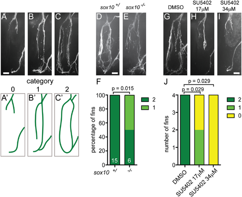
Nerve regrowth at the fin base requires sox10 and FGF. (A-C) Quantification of fin nerve regrowth in Tg(mnx1:GFP) transgenic larvae 24 hours after fin removal; nerves show different degrees of nerve regrowth (A-C). Categories 0, 1, and 2 indicate that none (A, A'), one (B, B'), or both sides (C,C') of the ring-like nerve network at the fin base regrew, respectively. (A'-C'): schematic representation of these results. (D-F) Nerve regrowth at the fin base 24 hours after fin removal in a wild type sibling (D) and a sox10 mutant (E). Sox10 mutants display significantly reduced nerve regrowth compared to wild type siblings. Graphical representation of the extent of nerve regrowth (category 0, 1, 2; F). (G-J) Nerve regrowth at the fin base at 24 hours after fin removal in a control larva treated with DMSO (G) and larvae treated with a low (17μM, H) or a high dose (34μM, I) of the FGF inhibitor SU5402. DMSO or SU5402 were added immediately after fin amputation. FGF inhibitor-treated larvae show significantly less nerve regrowth compared to DMSO-treated controls. Graphical representation of the extent of nerve regrowth (category 0, 1, 2; J). All scale bars are 10 μm. |