
Fig. 4
- ID
- ZDB-FIG-171016-9
- Publication
- Hübner et al., 2017 - Wnt Signaling Positively Regulates Endothelial Cell Fate Specification in the Fli1a-Positive Progenitor Population via Lef1
- Other Figures
- All Figure Page
- Back to All Figure Page
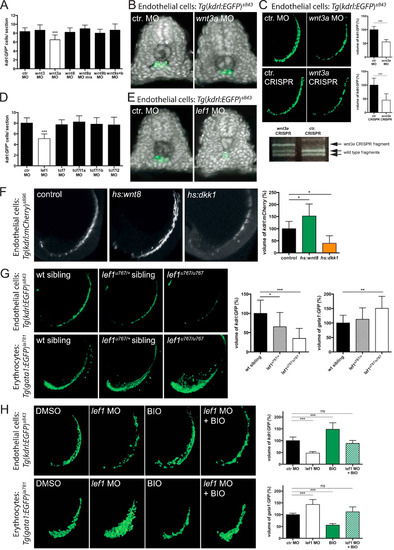
EC specification is induced by Wnt3a and mediated via Lef1. (A,B) Morpholino (MO)-mediated knockdown of wnt3a reduced the EC number (A, n = 14) in Tg(kdrl:GFP)s843 embryos compared to control (ctr.) MO (A, n = 19). Knockdown of other Wnt ligands, expressed in the zebrafish posterior LPM, did not significantly change EC numbers compared to ctr. MO injected embryos (A): wnt9a (n = 13), wnt9b (n = 14), wnt8 (n = 19) or wnt3 (n = 7). (C) MO-mediated knockdown or transient CRISPR-Cas9-mediated knockdown of wnt3a significantly reduced the EC volume in Tg(kdrl:GFP)s843 embryos (ctr. MO: n = 5; wnt3a MO: n = 8; ctr. CRISPR: n = 5; wnt3a CRISPR: n = 8). Agarose gel electrophoresis> of wnt3a CRISPR genotyping showed an undigested putatively mutated PCR product (wnt3a CRISPR fragment). Non-mutated wild type fragments were cut by PstI digest. (D,E) MO-mediated knockdown of lef1 reduced the EC number (D, n = 10) compared to ctr. MO (D, n = 38) in Tg(kdrl:GFP)s843 embryos. Knockdown of any of the other Tcf-transcription factors did not significantly change EC numbers compared to ctr. MO injected embryos (D): tcf7 (n = 30), tcf7l1a (n = 23), tcf7l1b (n = 28) or tcf7l2 (n = 32). (F) Heatshock induced Wnt ligand overexpression using Tg(hsp70l:wnt8-GFP)w34 (indicated as hs:wnt8) increased the EC volume in Tg(kdrl:mCherry)s896 embryos (F, n = 7) compared to control siblings (F, n = 12). In contrast, heatshock induced overexpression of Wnt signaling inhibitor Dkk1 using Tg(hsp70l:dkk1-GFP)w32 (indicated as hs:dkk1) decreased the EC volume (F, n = 5) compared to control siblings. (G) EC and erythrocyte cell volume analysis in lef1u767 mutant embryos. Embryos carrying heterozygous and homozygous lef1u767 mutations showed reduced EC volumes (G, upper panel, wt sibling: n = 14; lef1u767/+: n = 18; lef1u767/u767: n = 10), but increased erythrocyte volumes (G, lower panel, wt sibling: n = 14; lef1u767/+: n = 19; lef1u767/u767: n = 9) compared to wild type siblings. (H) MO-mediated knockdown of lef1 decreased the EC volume (H, upper panel, n = 6) compared to ctr. MO (H, upper panel, n = 7) in Tg(kdrl:GFP)s843 embryos, but increased the erythrocyte volume (H, lower panel, n = 7) compared to ctr. MO (H, lower panel, n = 7) in Tg(gata1:GFP)la781 embryos. Overactivation of Wnt signaling in lef1-deficient embryos using BIO restored EC volume and erythrocyte volume nearly to control levels (H, upper and lower panel, n = 7). All values represent mean±SD. *p<0.05, **p<0.01, ***p<0.001; Student's t-test. |
Reprinted from Developmental Biology, 430(1), Hübner, K., Grassme, K.S., Rao, J., Wenke, N.K., Zimmer, C.L., Korte, L., Mu Ller, K., Sumanas, S., Greber, B., Herzog, W., Wnt Signaling Positively Regulates Endothelial Cell Fate Specification in the Fli1a-Positive Progenitor Population via Lef1, 142-155, Copyright (2017) with permission from Elsevier. Full text @ Dev. Biol.