
Fig. 2
- ID
- ZDB-FIG-171013-19
- Publication
- Hübner et al., 2017 - Wnt Signaling Positively Regulates Endothelial Cell Fate Specification in the Fli1a-Positive Progenitor Population via Lef1
- Other Figures
- All Figure Page
- Back to All Figure Page
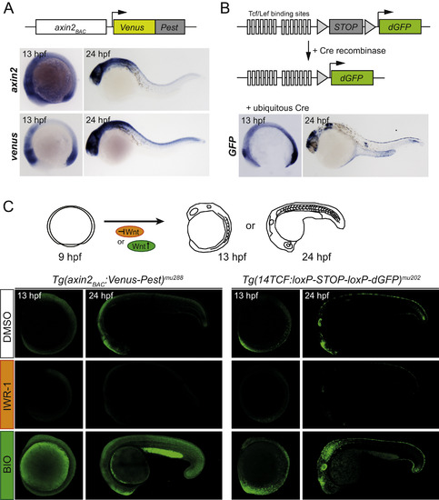
Tg(axin2BAC:Venus-Pest)mu288 and Tg(14TCF:loxP-STOP-loxP-dGFP)mu202 lines represent bona fide β-catenin dependent Wnt signaling reporters. (A,B) Domain structure of Tg(axin2BAC:Venus-Pest)mu288 (A) and Tg(14TCF:loxP-STOP-loxP-dGFP)mu202 (B), and whole mount in situ hybridization using antisense axin2, Venus and GFP probes. axin2 and Venus and GFP are expressed in similar domains: at 13 hpf hindbrain-midbrain boundary, posterior neuroectoderm and mesoderm; at 24 hpf in parts of the developing CNS, the skin and slightly in the tail tip with the GFP expression at 24hpf being limited to smaller domains in the CNS. (C) Tg(axin2BAC:Venus-Pest)mu288 (left panel) and Tg(14TCF:loxP-STOP-loxP-dGFP)mu202 (right panel) embryos were exposed to a Wnt signaling inhibitor (IWR-1) or a Wnt signaling activator (BIO) from 9 to 13 hpf or to 24 hpf and analyzed by confocal microscopy. Treatment with IWR-1 strongly reduced the fluorescence signal of both reporter lines compared to DMSO controls. Vice versa, treatment with Wnt signaling activator BIO resulted in strongly increased expression of both reporters compared to the respective controls. Hence, Tg(axin2BAC:Venus-Pest)mu288 and Tg(14TCF:loxP-STOP-loxP-dGFP)mu202 faithfully respond to Wnt signaling stimulation. For analysis, Tg(14TCF:loxP-STOP-loxP-dGFP)mu202 embryos were injected with cre mRNA at single cell stage. |
Reprinted from Developmental Biology, 430(1), Hübner, K., Grassme, K.S., Rao, J., Wenke, N.K., Zimmer, C.L., Korte, L., Mu Ller, K., Sumanas, S., Greber, B., Herzog, W., Wnt Signaling Positively Regulates Endothelial Cell Fate Specification in the Fli1a-Positive Progenitor Population via Lef1, 142-155, Copyright (2017) with permission from Elsevier. Full text @ Dev. Biol.