
Fig. 1
- ID
- ZDB-FIG-171006-2
- Publication
- Matsui et al., 2017 - An optimized method for counting dopaminergic neurons in zebrafish
- Other Figures
- All Figure Page
- Back to All Figure Page
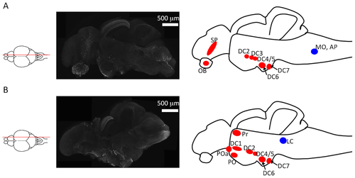
Identification of tyrosine hydroxylase (TH)+ neurons in sagittal sections of zebrafish brains. (A) Left panel: an illustration of the sectioning position; middle panel: TH+ immunoreactivity in a microsliced section of a 6-month-old zebrafish brain; right panel: TH+ neuron clusters found in this section. The red and blue circles indicate dopaminergic and noradrenergic neurons, respectively. (B) Left panel: an illustration indicating the sectioning position. The middle panel is an immunostaining of TH using a microsliced section of 6-month-old zebrafish. The right panel illustrates the TH+ neuron clusters in this section. The red and blue circles indicate dopaminergic and noradrenergic neurons, respectively. DC1–7: diencephalic catecholaminergic cluster. Abbreviations: AP, area postrema; LC, locus coeruleus; MO, medulla oblongata interfascicular zone and vagal area; OB, olfactory bulb; PO, preoptic region; POa, anterior preoptic region; Pr, dorsal pretectum; SP, subpallium. |
| Antibody: | |
|---|---|
| Fish: | |
| Anatomical Terms: | |
| Stage: | Adult |