
Fig. 4
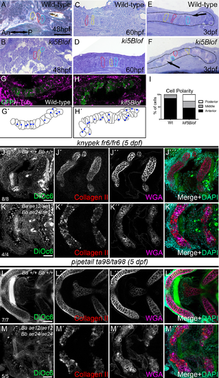
kif5Blof defects and interactions with Wnt PCP components. A-F) Semi-thin sections of the trabecular (A, B) and palatoquadrate cartilage (C-F). No differences were detected at 48 hpf (A, B) or 60 hpf (C, D). At 3 dpf, in Wt (E) chondrocytes stack like coins and span the cartilage. However, kif5Blof chondrocytes are circular (F), and close to the cartilage border. Some cells are delineated with dashed outlines and in E-F the cartilage limit is drawn. G-I) Analysis of MTOC orientation using γ-Tubulin. In Wt embryos (G, G´) >80% were anteriorly oriented (I). MTOC position was random in kif5Blof mutants (H, H'). WT: n = 8; 72cells. kif5Blof: n = 5; 63 cells (I). J-K''') Confocal images of single kny (J-J''') or triple mutants (K-K''') at 5 dpf. Secretion is normal in single kny mutants (J'-J''') but stacking is defective (J'''). Stacking and secretion defects in triple mutants (K-K'''). L-M''') Confocal images of single ppt (L-L''') or triple mutants (M-M'''). Secretion is intact in ppt single mutants (L'-L''') but stacking is disrupted (L'''). Membrane (M), secretion (M', M'') and stacking (M''') deficits are evident in triple mutants. ch: ceratohyal; m: Meckel´s; pq: palatoquadrate. Scale bars: A-F: 50μm; G, H: 20μm, J-M''': 50μm. |
| Gene: | |
|---|---|
| Antibodies: | |
| Fish: | |
| Anatomical Terms: | |
| Stage Range: | Protruding-mouth to Day 5 |
| Fish: | |
|---|---|
| Observed In: | |
| Stage Range: | Protruding-mouth to Day 5 |