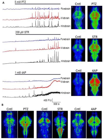
Fig. S4
- ID
- ZDB-FIG-170906-4
- Publication
- Winter et al., 2017 - 4-dimensional functional profiling in the convulsant-treated larval zebrafish brain
- Other Figures
- All Figure Page
- Back to All Figure Page
|
Wide-field imaging reveals CNS region-specific convulsant induced hyperactivity A) Left, time courses of GCaMP fluorescence intensity from time series collected with wide-field fluorescence imaging in three typical experiments. The bars indicate the time of drug application for PTZ, Strychnine and 4-AP and the portion of the trace to the left hand side of these bars indicates the pre-treatment (baseline) level of activity. The basal level of fluorescence was ~200 fluorescence intensity units in each area and the traces have been offset for clarity. Right, mean intensity projections across 300 consecutive frames (i.e. 5 min) collected either just prior to drug addition (left), or after 40-45 minutes drug treatment (right). The upper left image illustrates where the forebrain, midbrain and hindbrain regions of interest were located. The corresponding traces are coloured blue, red and black in each of the time course plots. B) Plots to indicate the degree of signal variance seen across the imaged x-y plane. These were compiled by creating projections of standard deviation across 300 frames and then (because SD is higher in the drug treated time-points) normalizing each pixel to the average standard deviation across the entire CNS. These normalized images were then pseudo-coloured and displayed between 1/3rd of mean SD (blue) and 3 times mean SD (red). Note how the drugs cause areas of greatest variation to change relative to their in subject controls and also how the different drugs seem to create different patterns.,/p> |

