
Fig. 3
- ID
- ZDB-FIG-170830-20
- Publication
- Xia et al., 2017 - Zebrafish slc30a10 deficiency revealed a novel compensatory mechanism of Atp2c1 in maintaining manganese homeostasis
- Other Figures
- All Figure Page
- Back to All Figure Page
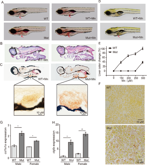
Slc30a10 mutant zebrafish develop liver damage. (A) Oil red O staining shows that mutant embryos have a slightly fatty liver (red arrows), which was worsened upon exposure to Mn. (B-C) HE staining (B) and Oil red O staining (C) of frozen sections showing hepatic steatosis in mutant embryos following Mn exposure. (D) Example images of a wild-type and mutant embryo under Mn exposure, showing a darker colored liver in the mutant (red arrow). (E) Dose-response curve showing the percentage of embryos with liver color change versus Mn concentration (n = 3 sets of 20 embryos/group). (F) Image of the liver of a wild-type and mutant adult, showing severe fibrosis in the mutant liver with sirius red staining. (G-H) The fibrosis markers col1a1a and ctgfa were measured in the liver of both male and female WT and mutant animals (n = 3 sets of 20 adults/group; *p<0.05 and **p<0.01). |
| Genes: | |
|---|---|
| Fish: | |
| Condition: | |
| Anatomical Term: | |
| Stage: | Adult |
| Fish: | |
|---|---|
| Condition: | |
| Observed In: | |
| Stage Range: | Day 6 to Adult |