
Fig. S6
- ID
- ZDB-FIG-170809-17
- Publication
- Mesureur et al., 2017 - Macrophages, but not neutrophils, are critical for proliferation of Burkholderia cenocepacia and ensuing host-damaging inflammation
- Other Figures
- All Figure Page
- Back to All Figure Page
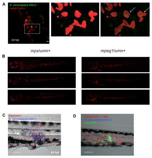
Related to Figs 6 and 7. Macrophages, but not neutrophils, contribute to increased bacterial burden and pro-inflammatory responses towards subcutaneous B. cenocepacia. (A) mpeg/umn+ embryo infected subcutaneously at 2 dpf with B. cenocepacia K56-2 (green). Confocal stack (59 slices, 1 μm) at 24 hpi showing bacteria in macrophages. A close up of the indicated area is shown in 3D, with volume rendering for GFP signal and surface rendering of mCherry signal without (middle panel) and with 50% surface transparency (right panel). Scale bar 50 μm. (B) Fluorescence images showing non-infected mpx/umn+ and mpeg1/umn+ control embryos at 3 dpf, the time point that resembles 24 h post subcutaneous injection in Fig 6. (C) Image overlay (bright field, red and blue filters) of the tail region of an mpx/umn+ embryo after subcutaneous injection with B. cenocepacia K56-2 expressing Turquoise, presenting tissue damage by 24 hpi. Scale bar, 100 μm. (D) Image overlay (bright field, red, blue and green filters) of the tail region of an Mtz-treated mpeg1/umn+; mpx:GFP embryo 24 h after subcutaneous injection with B. cenocepacia K56-2 expressing Turquoise. Scale bar, 100 μm. |