
Fig. 6
- ID
- ZDB-FIG-170807-26
- Publication
- Passoni et al., 2017 - Imaging of viral neuroinvasion in the zebrafish reveals that Sindbis and chikungunya viruses favour different entry routes
- Other Figures
- All Figure Page
- Back to All Figure Page
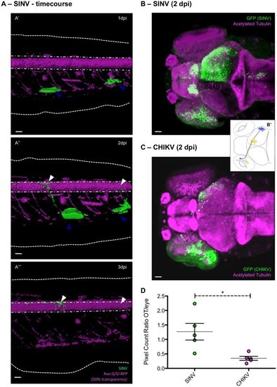
Efficient axonal transport of SINV. (A) Infection of muscle cells and connected spinal cord neurons in the tail region of a huc:G/U:RFP larva. Live confocal imaging, maximal projection, 3D rendering, with superposition of green (infected cells) and magenta (neurons) fluorescence. Same larva imaged at 1 (A′), 2 (A″) and 3 dpi (A‴). Dotted white lines indicate the limits of the fins; dash-dotted lines the limits of the spinal cord. Scale bars: 50 µm. (B,C) Assay for axonal transport to the contralateral optic tectum after inoculation of SINV (B) or CHIKV (C) in the left retina. Confocal imaging of fixed infected larva at 2 dpi, with superposition of green (infected cells) and magenta (acetylated tubulin) fluorescence; maximal projections. Scale bars: 50 μm. (B′) Scheme of the projection of the retinal neurons to the optic tectum. (D) Ratio of GFP fluorescence intensities measured in the eye and the contralateral optic tectum, after SINV or CHIKV inoculation. n=5 from two independent experiments pooled. |
| Gene: | |
|---|---|
| Antibody: | |
| Fish: | |
| Conditions: | |
| Anatomical Terms: | |
| Stage Range: | Day 4 to Day 6 |