
FIGURE
Fig. 3
Fig. 3
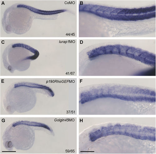
Expression pattern of muscle-specific mhc in representative control and morphant embryos at 24 hpf. (A,B) A CoMO-injected embryo showing regular mhc expression pattern in the somites. (C,D) A lurap1MO-injected embryo with bent axis and disrupted mhc expression pattern in the somites. (E,F) A p190RhoGEFMO-injected embryo with shortened anteroposterior axis and absence of mhc expression at somite boundaries. (G,H) A Golgin45MO-injected embryo with reduced anteroposterior axis associated with disrupted mhc expression pattern in the somites. Scale bars: (A,C,E,G) 350 μm; (B,D,F,H) 120 μm. |
Expression Data
Expression Detail
Antibody Labeling
Phenotype Data
| Fish: | |
|---|---|
| Knockdown Reagents: | |
| Observed In: | |
| Stage: | Prim-5 |
Phenotype Detail
Acknowledgments
This image is the copyrighted work of the attributed author or publisher, and
ZFIN has permission only to display this image to its users.
Additional permissions should be obtained from the applicable author or publisher of the image.
Full text @ Sci. Rep.