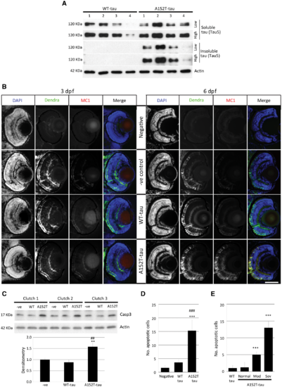
Tau aggregation and cell death in Dendra-tau transgenic zebrafish. (A) Levels of sarkosyl-soluble and insoluble tau reflect accumulation of the insoluble form only in A152T-tau fish at 6 dpf. The level of total tau was analysed by immunoblotting using Tau5 antibody (four independent clutches for WT-tau and A152T-tau). (B) Antibody staining for the conformational marker MC1 in cryosections across the eye of WT-tau and A152T-tau fish. No staining was observed in either WT-tau or A152T-tau fish at 3 dpf (left), whereas only A152T-tau presented positive staining for conformational changes at 6 dpf (right). Scale bar = 100 μm. (C) Western blot for active Caspase 3 (Casp3) (quantified below), indicative of increased cell death in fish expressing the A152T variant (mean ± SEM of nine independent clutches; Student-Newman-Keuls one-way ANOVA, *P < 0.01 versus negative, ##P < 0.01 versus WT-tau). (D) The increased cell death in A152T-tau fish was confirmed by quantification of TUNEL labelling on transverse sections (mean ± SD; n = 5 fish, from a minimum of five sections; Student-Newman-Keuls one-way ANOVA, ***P < 0.001 versus negative; ###P < 0.001 versus WT-tau). See also Supplementary Fig. 4. (E) Morphologically abnormal A152T-tau fish showed increased cell death (quantification of TUNEL-positive nuclei) compared to morphologically normal A152T- or WT-tau fish (mean ± SD; Student-Newman-Keuls one-way ANOVA, **P < 0.01 and ***P < 0.001 versus WT-tau). Representative images in Supplementary Fig. 7.
|
