
Fig. S2
- ID
- ZDB-FIG-170517-27
- Publication
- Kaslin et al., 2017 - Distinct roles of neuroepithelial-like and radial glia-like progenitor cells in cerebellar regeneration
- Other Figures
- All Figure Page
- Back to All Figure Page
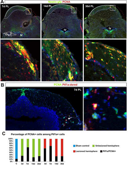
A.Confocal maximum projections of cerebellar cross sections showing stem cell activation after injury. nestin:gfp+ (green) neuroepihelial-like stem cells and proliferating cells labelled with PCNA (red). B. Confocal maximum projections of cerebellar cross sections showing a proliferating (green) ptf1a:Dsred+ (red) VZ progenitor in the parenchyma. Proliferating cells labelled with PCNA (green), DAPI (blue). C. Quantification of proliferating cells among the pool of Dsred+ cells. Dsred is very stable (many days) in the cells and persist in differentiating cells. The proportion of PCNA+ cells is reduced notably 14 days after injury suggesting that majority of Dsred+ cells are differentiating. (Sham control n=7, 3DPL n=4, 7DPL n=5, 14DPL n=5, 30 DPL n=5). |