
Fig. S1
- ID
- ZDB-FIG-170517-26
- Publication
- Kaslin et al., 2017 - Distinct roles of neuroepithelial-like and radial glia-like progenitor cells in cerebellar regeneration
- Other Figures
- All Figure Page
- Back to All Figure Page
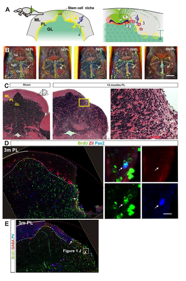
A. Overview of the adult stem cell niche in the cerebellum. Stem cells are located around a small out pocketing of the fourth ventricle. 1. Polarized neuroepithelial-like stem cells (green) are restricted to the midline of the dorsal cerebellum. The stem cells give rise to rapidly migrating granule precursors (dark green) that initially migrate dorsolaterally and give rise to granule neurons in a distinct outside-in fashion. 2. Radial glia-like stem and progenitor cells are located ventral to the neuroepithelial-like stem cells and largely quiescent in the adult. 3. Bergmann glia and inhibitory inter-neurons are produced at a low level at the lateral margin of the ventricle. In addition, rare stem cells reside in the cerebellar parenchyma. GL=granule cell layer, ML=molecular cell layer, PL=Purkinje cell layer. B. Representative timelapse of a lesioned fish showing rapid wound healing after injury. Dorsal view of the lesion site (hatched white line and white arrow). The injury is fully covered three days after injury and at five days after injury pigment cells are readily detected at the injury site. Ce=Cerebellum, PL=post lesion, Tel=Telencephalon, TeO=Optic tectum. C. Hematoxylin and Eosin stained cerebellar cross section from sham injured and injured fish twelve month after injury (n=5/condition). The Purkinje and molecular cell layer has recovered very poorly. No notable fibrotic or glial scarring is detected. GL=granule cell layer, ML=molecular cell layer, PL=Purkinje cell layer. D. Cerebellar cross section showing BrdU (green), ZebrinII (Red) and Pax2 (Blue) staining three months after injury. No BrdU/ZII positive cells are detected and very few BrdU/Pax2 positive cells are detected (arrow). E. Cerebellar cross section showing BrdU (green), GABA (Red) and Parvalbumin (Blue) staining three months after injury. No BrdU/PV positive cells are detected and very few BrdU/GABA positive cells are detected (arrows). Yellow hatched line show original picture border in rotated in images. |